
 By
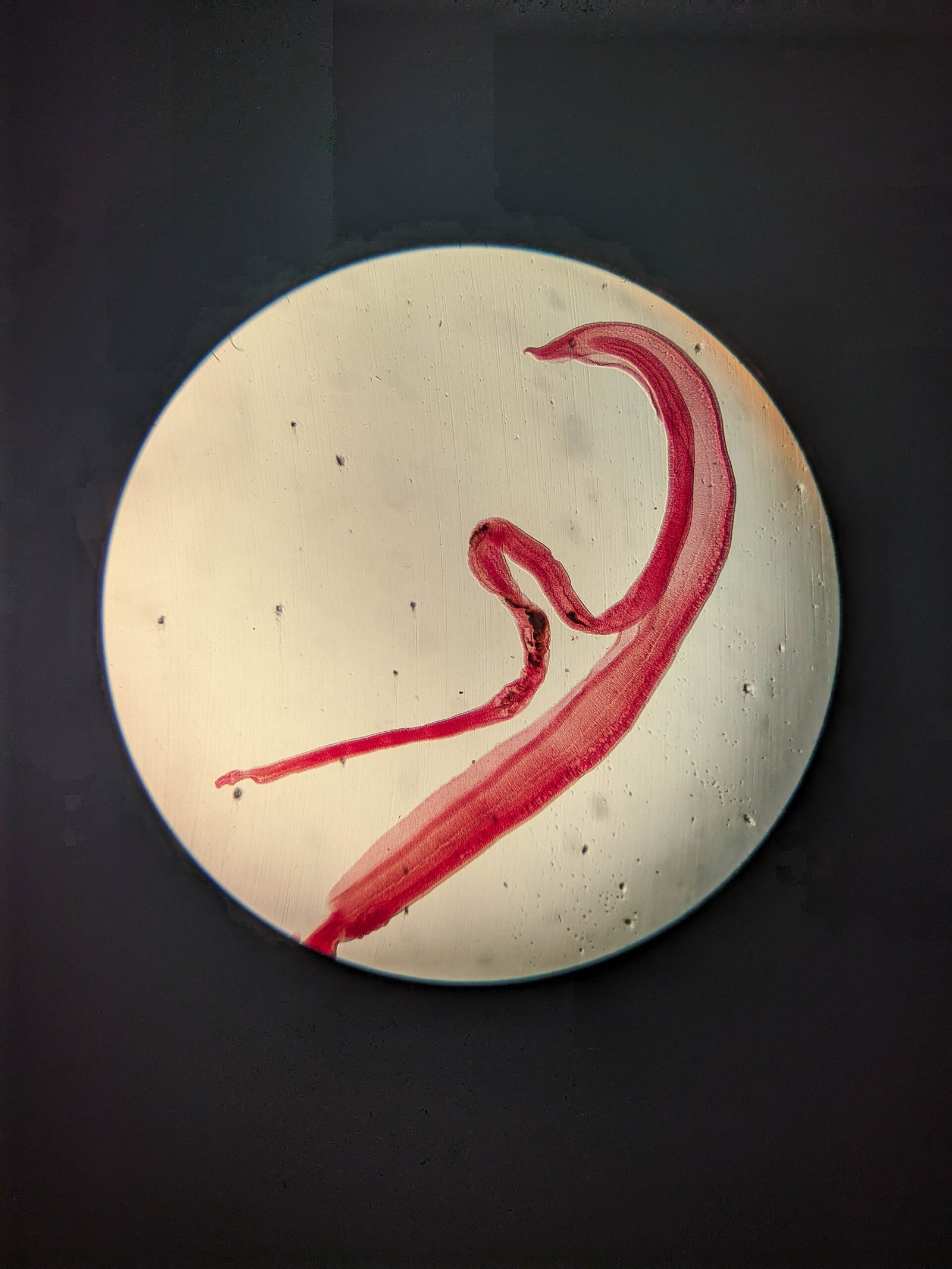
By In an extraordinary intersection of clinical neurology and advanced parasitology, medical specialists have successfully confirmed a rare parasitic brain infection in a patient following an unprecedented laboratory breakthrough. Because no single, definitive biomarker or standardized blood test currently exists to verify the presence of this specific pathogen within human central nervous tissue, clinical teams relied on specialized microbiological culturing. The breakthrough occurred when microscopic eggs isolated during diagnostic evaluation successfully germinated in vitro, developing unmistakable caudal tails that provided undeniable structural proof of the organism’s identity.
Strategic Background & Clinical Timeline
Central nervous system parasitic infections represent some of the most challenging conditions in contemporary medicine. Patients frequently present with nonspecific neurological symptoms, ranging from persistent cranial pressure and unexplained cognitive deficits to localized motor dysfunction. In this case, the patient underwent months of extensive medical evaluations, including high-resolution magnetic resonance imaging (MRI), cerebral spinal fluid analysis, and broad-spectrum infectious disease screening, all of which yielded inconclusive or ambiguous results.
Standard serological testing for neuroparasitic agents often suffers from high rates of cross-reactivity or false negatives, particularly when the pathogen is localized within the immune-privileged sanctuary of the brain. Recognizing the limitations of standard diagnostic panels, treating physicians collaborated with senior diagnostic microbiologists to isolate biological samples for specialized microscopic culture. Over an extended incubation window, laboratory specialists maintained rigid environmental parameters mimicking human biological tissue, ultimately observing the morphological transformation of encapsulated eggs into motile, tail-bearing larvae.
Q&A Roundup: Unraveling the Diagnostic Breakthrough
Q: Why are central nervous system parasitic infections notoriously difficult to diagnose using standard medical technologies?
A: Diagnosing parasitic invasions of the human brain poses a monumental clinical challenge due to several physiological and technological hurdles. First, the blood-brain barrier severely limits the concentration of systemic antibodies that enter the cerebrospinal fluid, rendering standard blood and spinal fluid antibody tests far less sensitive. Second, advanced neuroimaging tools like CT and MRI scans can readily detect tissue lesions, inflammation, or cystic structures, but they cannot definitively differentiate between parasitic cysts, primary brain tumors, fungal granulomas, or autoimmune lesions. Without a direct tissue sample or a unique biological marker, clinicians are often left with indirect clues rather than definitive diagnoses.
Q: What specific event in the laboratory provided the definitive diagnostic confirmation?
A: The critical breakthrough occurred within the controlled environment of the diagnostic microbiology laboratory. Having isolated microscopic ovum-like structures from clinical specimens, microbiologists placed the samples into specialized nutrient media designed to support delicate organism viability. After several days of precise thermal and oxygenation control, microscopic analysis revealed that the eggs were not only viable but were actively undergoing morphogenesis. The critical visual proof came when the developing embryos sprouted distinct, articulated caudal tails—a defining morphological characteristic of this specific parasitic lineage. This phenotypic transformation provided the indisputable physical evidence that modern molecular screening had failed to deliver.
Q: How does this case highlight current gaps in biotechnology and laboratory diagnostics?
A: This landmark case exposes a critical gap in contemporary diagnostic biotechnology: our reliance on automated immunoassays and genetic sequencing protocols that require prior reference genomes. When dealing with rare, mutated, or under-researched parasitic strains, standard polymerase chain reaction (PCR) assays can fail if the primer sequences do not perfectly match the organism’s genetic code. The successful identification in this instance relied on classic, observational microbiology augmented by modern environmental culturing, proving that functional phenotypic observation remains an indispensable weapon in the diagnostic arsenal when molecular assays fall short.
Without a single definitive biomarker or serological standard for this specific parasite, the microscopic observation of motile tail development within controlled laboratory cultures provided the unmistakable visual confirmation our clinical teams desperately needed.
Economic & Global Health Implications
The success of this diagnostic procedure carries significant implications for clinical research funding, global health monitoring, and biotechnological development. As international travel, environmental shifts, and changing ecological habitats alter the geographic distribution of rare pathogens, urban medical centers across the globe are increasingly encountering exotic parasitic conditions previously restricted to specific tropical or subtropical regions.
Healthcare systems face growing economic pressures to streamline diagnostic pipelines for rare infectious diseases. Prolonged diagnostic uncertainty results in repeated hospitalizations, unnecessary invasive procedures, and extended empirical treatment regimens that carry substantial financial costs and patient risks. Investing in integrated diagnostic laboratories capable of performing both high-throughput molecular sequencing and traditional live-culture morphology is proving essential for modern medical infrastructure.
Key Takeaways & Strategic Intelligence
- Definitive Morphological Proof: Confirmation of the central nervous system infection was achieved after isolated parasitic eggs developed visible caudal tails in vitro, establishing absolute etiology.
- Limitations of Conventional Assays: Traditional neuroimaging and broad-spectrum blood panels proved inconclusive, underscoring the systemic difficulty of detecting brain parasites without physical pathogen culture.
- Methodological Synthesis: The case demonstrates the necessary synergy between advanced biological incubators and fundamental observational microbiology in modern diagnostic medicine.
- Global Vector Expansion: Healthcare providers must prepare for an increased prevalence of atypical parasitic presentations driven by changing global travel patterns and environmental shifts.
Future Outlook & Strategic Projections
Looking ahead, medical technology firms and research institutions are expected to accelerate the development of microfluidic bio-chips and artificial intelligence-enhanced microscopic vision systems capable of rapidly recognizing larval morphological changes in real time. Furthermore, this landmark case is likely to catalyze renewed academic interest in neuroparasitology diagnostic standards, encouraging regulatory bodies to establish standardized protocols for live-culture monitoring when conventional molecular and radiological tests yield equivocal results. For the patient, definitive identification enables targeted anti-parasitic therapeutics, shifting the clinical focus from uncertain diagnostic management to structured recovery and neurological rehabilitation.